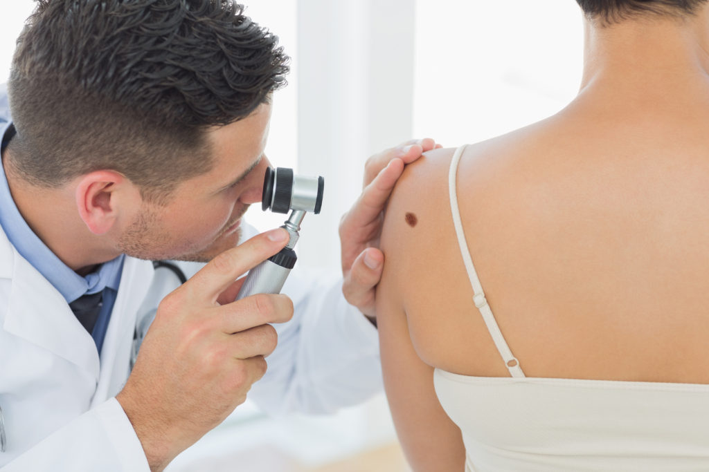
With Australia and North QLD having the dubious honour of leading the world in the incidence of skin cancer including melanoma, Whitsunday Family Practice prioritises providing the latest technology and services to the doctors using our facility. Approximately two in three Australians will be diagnosed with skin cancer by the time they are 70, with more than 750,000 people treated for one or more non-melanoma skin cancers each year.
Our clinic facilities provide doctors with the use of the latest Dermoscopy equipment for skin checks – a handheld dermoscope device that uses polarised light and magnification to assist the doctor when observing coloured skin lesions in the deeper skin layers, increasing the chance of detecting melanoma.
In addition, our practice provides the facility Dermengine – a leading digital platform to compare and analyse skin lesions and allow accurate monitoring of suspect lesions over a period of time.
Whilst all of the doctors providing services are able to perform skin checks and excisions of lesions, Dr Dylan MacLeod, Dr May Oo, Dr Rose Si, Dr Khin Htet and Dr Melissa McCann have all completed additional training in performing skin checks and skin cancer management- Call us to book your skin check today on 074948 3323 or book online at https://www.whitsundayfamilypractice.com.au/bookings/
The following information is provided by Dr McCann for patients wishing to learn more about skin cancer-
This section helps to identify changes that might signal melanoma. Please see your doctor immediately if you have a skin lesion that you are worried about.
There is one universal truth about melanoma, and that is change.
Further to monitoring your moles for change, the ABCD and EFG rules will help you identify melanoma.
The A.B.C.D. rule has been very effective at aiding the early identification of superficial spreading melanomas. Superficial spreading melanoma can have any one of the following criteria:
ASYMMETRY: The shape of one half does not match the other.
There is a class of rapidly growing, nodular melanoma, which represents about 20% of all cases of melanoma. This type of melanoma does not subscribe to the ABCD rule and thus can go undetected. Fortunately, they do behave in a way that allows them to be identified early using the E.F.G. rule. Nodular melanoma usually has all three of the below criteria:
If you notice a lesion that is:
You should bring it to the attention of your doctor immediately, accurately describing the symptoms and your reason for concern.
This section contains information about these types of cancers, what to look for, and how to prevent them.
s the most common form of skin cancer. It occurs most frequently on sun-exposed regions of the body.
Although this skin cancer rarely spreads (metastasises) to other organs of the body, it can cause destroy surrounding tissue and disfigure the skin surface. So early detection and treatment are essential. Most basal cell carcinomas are caused by chronic sun exposure, especially in people with fair skin, light hair and blue, green or grey eyes. In a few instances, there are other contributing factors such as having had a previous BCC, burns, exposure to radiation or chronic dermatitis.
Basal cell carcinoma may have several different appearances on your skin. Some warning signs that may indicate basal cell carcinoma are an open sore, a reddish patch, a growth with an elevated border and a central indentation, a bump or nodule and a scar-like area.
Is a major type of cancer that arises from the outer epidermal layer of the skin and mucous membranes and occurs most commonly on areas exposed to the sun. If untreated, squamous cell carcinoma may penetrate and destroy underlying tissue. In a small percentage of cases, this tumour can spread (metastasise) to distant organs and may be fatal.
Chronic sun exposure is the leading cause of squamous cell carcinoma, especially in people with fair skin, light hair and blue, green or grey eyes. Other factors that may contribute to the development of this cancer include having had a previous SCC, burns, scars, chronic inflammatory conditions and immunosuppression.
Although more likely to develop in fair-skinned individuals, squamous cell carcinoma may occur in dark-skinned people, especially at sites of preexisting inflammation. Signs that may indicate the presence of squamous cell carcinoma include scaly red patches, elevated growth with a central depression, wart-like growths, nodules and open sores.
Is a very serious form of skin cancer of melanocytes, the cells that produce a dark protective pigment called melanin.
Individual lesions may appear as a dark brown, black or multi-coloured growth with irregular borders that can become crusted and bleed.
Melanoma may affect anyone at any age and can occur anywhere on the body, not only on sun-exposed areas.
An increased risk of developing this disease is seen in people who have fair skin, light hair and eye colour, a family history of melanoma or who have had melanoma in the past. These tumours can arise in or near a preexisting mole or may appear without warning.
Melanoma may spread to other organs, making it essential to treat this skin cancer early.
Further reliable information on skin cancer causes and treatment can be found at:
https://www.cancer.org.au/about-cancer/types-of-cancer/skin-cancer.html
There are various ways of managing skin cancer, with surgical removal remaining the first-line approach. Second-line options include topical creams and photodynamic therapy. Your doctor will discuss these with you as appropriate.
There are many other types of skin spots that are not cancerous, and the majority will be identified at your skin check. These are usually managed non-surgically, and various other management options are available within the clinic. These two particular types of lesions have a higher risk of becoming cancerous:
Also called atypical naevus or dysplastic naevus. These are benign moles that may share some of the clinical or microscopic features of melanoma, but is not a melanoma or any other form of cancer. However, the presence of atypical naevi may increase the risk of developing melanoma or be a marker for someone who is at risk of developing melanoma. This increased risk varies from very small for those with a single atypical nevus to higher for those with many.
Also known as solar keratosis, these are precancerous lesions of the epidermis (top layer of the skin) that arise through long-term exposure to sunlight, commonly on the face, lips, ears, neck, scalp, forearms and backs of hands. They are very frequently found in most parts of Australia.
Chronic sunlight exposure alters the keratinocytes (cells that make up the majority of the epidermis) and causes patches of skin to become scaly, rough, discoloured and sometimes tender to touch.
Some AK will eventually turn to skin cancer, so these lesions should not be ignored.